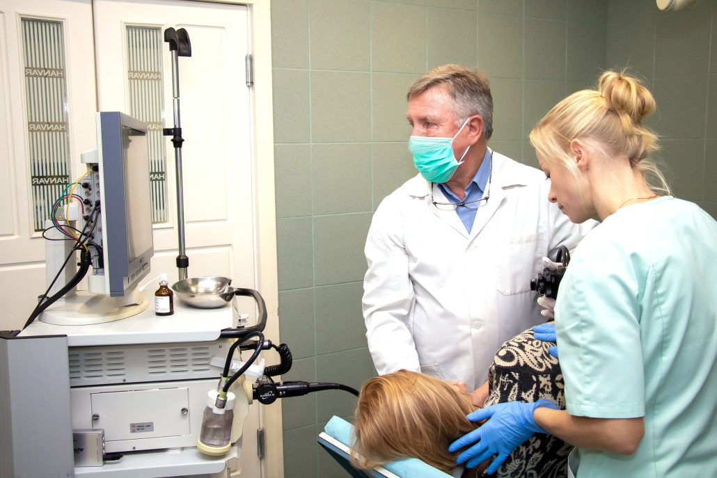
What is gastroscopy?
Gastroscopy is an examination of the inside of the gullet, stomach and duodenum. It is performed by using a thin, flexible fibre-optic instrument that is passed through the mouth and allows the doctor to see whether there is any damage to the lining of the oesophagus (gullet) or stomach, and whether there are any ulcers in the stomach or duodenum.
The GP will decide when drug treatment alone is sufficient or whether an investigation by gastroscopy at the local hospital is necessary.
The procedure is painless and is usually done under a light sedative as a day-case patient in a specialised endoscopy unit.
Occasionally, after a discussion with the endoscopist, the procedure will be performed without sedation. When sedation is used, the patient will not be able to drive or operate machinery for the rest of the day.
Anyone suffering from stomach problems should consult a doctor who will, in most cases, treat the symptoms without a major examination.
After explaining the procedure, the endoscopist will spray the back of the throat with a local anaesthetic. This is similar to the anaesthetic used by dentists. It numbs the throat and may make it difficult to swallow. When sedation is used, it is not a full anaesthetic and the patient will still be conscious and aware. A nurse will lie the patient on their left side and the endoscopist will then gently place the end of the instrument into the mouth and ask the patient to swallow it, which feels like swallowing a large piece of food.
The endoscopist may need to put some air into the stomach to perform the examination effectively and this can cause discomfort or even a need to belch. This is perfectly normal.
The endoscopist will closely examine the lining of the gullet, stomach and duodenum to identify the cause of the symptoms. It will take about 10 to 15 minutes
Why is gastroscopy useful?
- The doctor can study the mucous membrane of the stomach from the top to the bottom, and see irritation, wounds, or tumours. Gastroscopy is effective, and has now replaced the use of X-rays in many cases. It helps the doctor see any abnormalities in the gullet, the stomach and the duodenum. It is precise and safe.
- Through the gastroscope, the doctor can take samples or photographs of the mucous membrane. The most modern gastroscopes can also show the areas in the stomach on a TV screen, so that the mucous membrane can be studied thoroughly. This can be recorded on a videotape, and used for later comparison.
Patients are often given a gastroscopic examination because of their indigestion symptoms, which can usually be treated with tablets.
Occasionally, the cause of indigestion is an ulcer and it is now known that many ulcers are due to bacterial infection in the stomach.
A biopsy (a small piece of the lining of the stomach) may be removed during an endoscopy and examined under the microscope in the laboratory to pinpoint an infection.
A very small number of patients with indigestion will turn out to have cancer and, again, the diagnosis can be made accurately by biopsy. Further investigation can then be planned to ensure the most effective treatment.
How far can a gastroscope see?
A gastroscope can only examine the lining of the oesophagus (gullet) stomach and duodenum. It will detect conditions in those organs that are causing symptoms but will not, for example, detect gallstones or pancreatic disease.
All procedures carry some risk but outpatient diagnostic gastroscopy is very safe. Minor complications are uncommon and major complications are very rare.
What is colonoscopy? How is a gastroscopy performed? Is gastroscopy safe?
Colonoscopy is a test that allows your doctor to look at the inner lining of your large intestine (rectum and colon). He or she uses a thin, flexible tube called a colonoscope to look at the colon. A colonoscopy helps find ulcers, colon polyps, tumors, and areas of inflammation or bleeding. During a colonoscopy, tissue samples can be collected (biopsy) and abnormal growths can be taken out. Colonoscopy can also be used as a screening test to check for cancer or precancerous growths in the colon or rectum (polyps).
The colonoscope is a thin, flexible tube that ranges from 48 in. (122 cm) to 72 in. (183 cm) long. A small video camera is attached to the colonoscope so that your doctor can take pictures or video of the large intestine (colon). The colonoscope can be used to look at the whole colon and the lower part of the small intestine. A test called sigmoidoscopy shows only the rectum and the lower part of the colon.
Before this test, you will need to clean out your colon (colon prep). Colon prep takes 1 to 2 days, depending on which type of prep your doctor recommends. Some preps may be taken the evening before the test. For many people, the prep for a colonoscopy is more trying than the actual test. Plan to stay home during your prep time since you will need to use the bathroom often. The colon prep causes loose, frequent stools and diarrhea so that your colon will be empty for the test. The colon prep may be uncomfortable and you may feel hungry on the clear liquid diet. If you need to drink a special solution as part of your prep, be sure to have clear fruit juices or soft drinks to drink after the prep because the solution tastes salty.
Colonoscopy is one of many tests that may be used to screen for colon cancer. Which screening test you choose depends on your risk, your preference, and your doctor. Talk to your doctor about what puts you at risk and what test is best for you.


